
Medical professionals are turning to advanced technology to solve an expansive list of issues related to differentiating between cell types during hematology analysis. Artificial intelligence and 3D imaging are cost-effective ways to classify unique biochemical traits in the blood. New techniques are emerging, which allow for high-speed imaging and mapping of live cell structures with imaging contrast. Let’s explore how 3D imaging is revolutionizing technology for hematology analyzers.
3D Imaging in Blood Analysis
Imaging gives hematology analyzers the ability to accurately map the thickness and density of blood. By acquiring visuals of living cells, the analyzer may extract biomarkers, measure cell growth, understand blood cell viability, and screen for disease. AI and advanced imaging enhance the blood analysis by allowing for cell classification and diagnosis. In fact, high-quality imaging creates unique models for identifying specific cell features and structures. These advances are essential to overcoming cell analysis obstacles inherent in less sophisticated hematology analyzers.
Artificial intelligence machine learning is an exciting and innovative development that incorporates bright and dark imaging to improve the accuracy of detecting blood abnormalities. As advancements in hematology analyzer technology continues, equipment is becoming more sophisticated. Handheld units offer on-the-spot evaluations in a physician’s office. Other advanced analyzers process as many as 100 blood samples every hour for busy labs. Using complex technology of hematology analyzers’ advanced optics and innovative software to deliver quick, efficient blood analysis, clinicians are incorporating 3D imaging into daily medical assessments.
Imaging Benefits
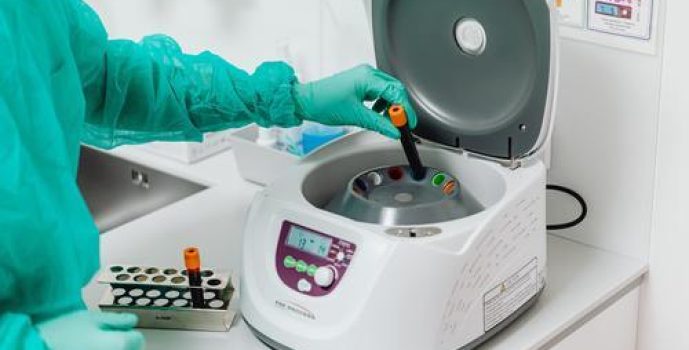
These 3D imaging techniques rely on a form of cytometric technology and optical flow to detect cell type and provide detailed information to clinicians. Advancing past traditional centrifugation techniques, imaging allows for accurate blood counts and allows for optical analysis. With flow cytometry high-speed and efficient analysis of blood cell populations is revolutionizing the way the medical profession assesses patient disease. In controlled environments, the hematology analyzers perform 3D output of biological models while providing high-definition blood cell resolution.
Complex blood analysis takes note of cell size, shape, and variations via the automated processes withing advanced hematology analyzers. As the alternative to less advanced systems, imaging cytometry performs multicell analysis and offers high technical capacity for identifying issues within the blood sample. Systems capable of analyzing complex cell development highlight the technology advancement in recent years, which goes beyond the most popular analysis techniques for blood cell analysis. Improvement in optical imaging for medical use has opened the door for a wide range of advancements in the field. By incorporating 3D imaging into hematology analyzers, manufacturers are improving patient care and assessment.


